Introduction
Nuclear medicine imaging devices detect and display the in vivo distribution of radiopharmaceuticals. Nuclear medicine imaging visualizes organs or lesions based on differences in radioactive concentration between normal and diseased tissues. Compared with CT and MRI, nuclear medicine imaging can detect and diagnose certain diseases at earlier stages. It is a functional imaging modality, also called radionuclide imaging.
1. Device Types and Characteristics
Gamma Camera
Composition:
- Scintillation probe: includes collimator, scintillation detector, and photomultiplier tubes.
- Electronics: includes preamplifier, single pulse height analyzer, and correction circuits.
- Display devices: oscilloscope, camera, and related equipment.
- Additional gamma camera accessories.
Characteristics:
- Continuous imaging can track and record the morphology and function of a radiopharmaceutical as it passes through an organ for dynamic studies.
- Relatively short examination time and operational simplicity make it suitable for pediatric and critically ill patients.
- Rapid imaging facilitates multi-position and multi-site observation.
- Image processing can produce diagnostic data and quantitative parameters.
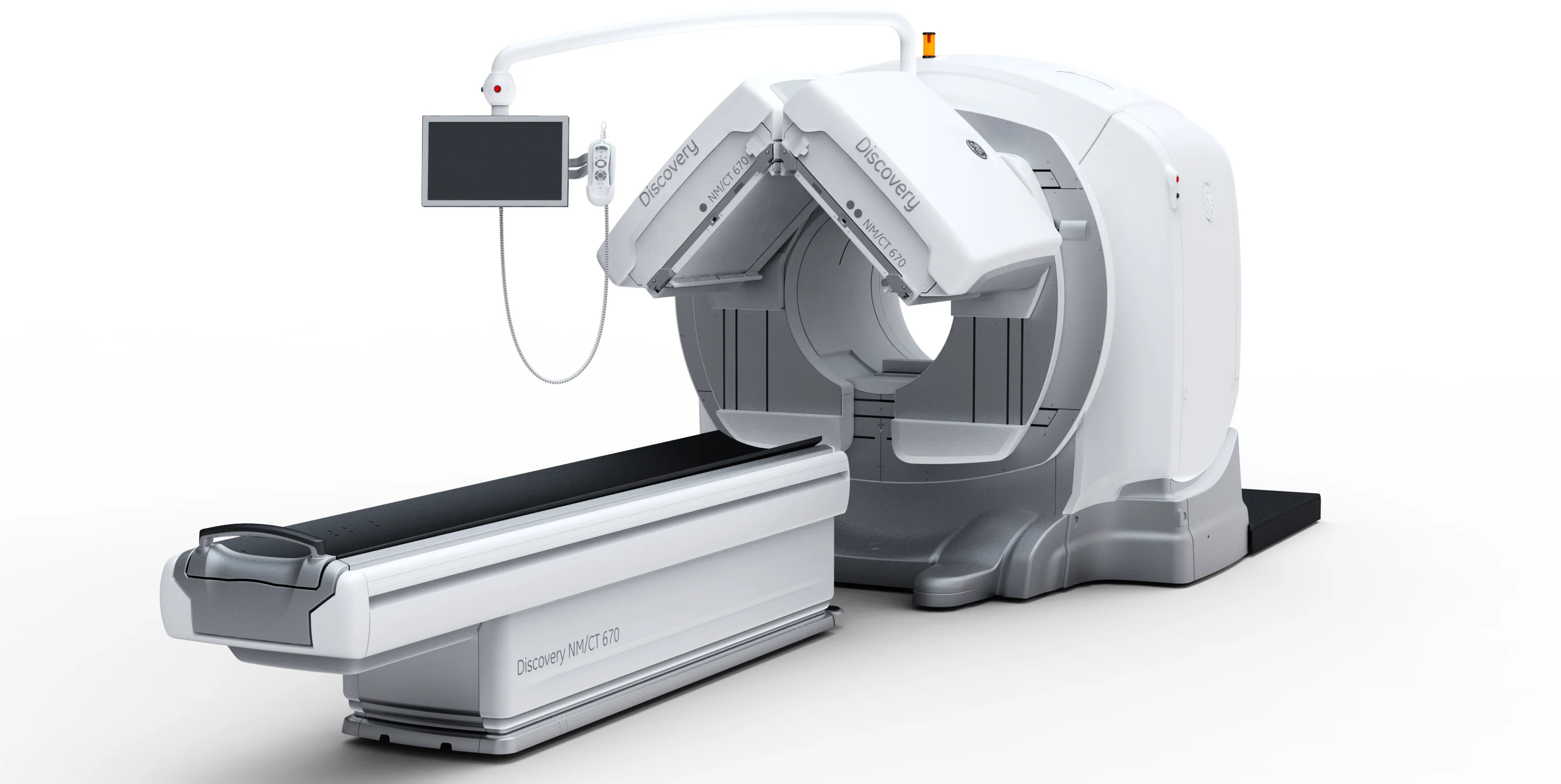
Single Photon Emission Computed Tomography (SPECT)
Imaging Principle
SPECT uses gamma cameras that rotate around the region of interest to collect emitted gamma photons at different angles. Counts from these projections are reconstructed using tomographic reconstruction algorithms similar to those used in X-CT to obtain the distribution of radiopharmaceutical concentration within a slice. This yields multi-planar slice images or a three-dimensional volume.
Typical SPECT systems measure photon energies from 50-600 keV and achieve spatial resolution of approximately 6-11 mm.
Differences from X-CT
- Coarser images and lower spatial resolution compared with X-CT.
- SPECT is an emission tomographic technique.
Positron Emission Tomography (PET)
PET uses positron-emitting radionuclides. Many biologically essential elements can be labeled with positron emitters without significantly altering their biological activity, allowing labeled compounds or metabolites to participate in physiological and biochemical processes. These radionuclides typically have short half-lives, permitting the use of higher administered activities to improve image contrast and spatial resolution. PET images therefore reflect physiological, biochemical, or pathological function.
Because positrons have a short range and annihilate almost immediately, they cannot penetrate thick tissue. PET detects the annihilation gamma photons resulting from positron-electron annihilation.
Limitations:
- Widespread PET use is constrained by two factors: the short half-life of positron-emitting radionuclides, which requires local production by a cyclotron, and the need for rapid radiopharmaceutical synthesis facilities and laboratories to prepare labeled tracers.
2. Imaging Process and Basic Requirements
- Label a pharmaceutical agent with a radioactive isotope to form a radiopharmaceutical and administer it to the patient. When the target organs or tissues absorb the agent, an internal radiation source is formed.
- External gamma detectors can measure the gamma rays emitted during radioactive decay and generate images that represent the in vivo distribution density of the radionuclide.
Because radiopharmaceuticals participate in normal metabolic processes like natural elements and other compounds, nuclear medicine images reflect not only organ morphology but, more importantly, organ function and related physiological and biochemical information.
3. Fundamental Characteristics of Nuclear Medicine Imaging
- Nuclear medicine imaging is based on radioactivity concentration differences within and between organs. It produces static and dynamic images that reflect the location, morphology, and size of tissues and lesions, and also provides information on regional and local functional changes.
- It supports various dynamic imaging modes. Organ uptake, absorption, and excretion of radiopharmaceuticals allow dynamic and quantitative visualization of blood flow and organ function, and provide functional parameters related to perfusion, metabolism, and receptor status.
- Certain radionuclides show specific accumulation in target organs or lesions, producing highly specific images useful for detecting different tumor types, receptor distributions, inflammation, and metastases—findings that are often difficult to obtain from morphology-based imaging alone.
4. Summary
Nuclear medicine is a developing discipline with distinct advantages. Imaging devices and diagnostic instruments have evolved alongside the field. From single-function measurement devices to integrated large-scale scanners, advances in nuclear medicine imaging equipment continue to support the clinical development of nuclear medicine.