Background
Global rates of pregnancy-related disorders are concerning. Each year there are more than 20,000,000 high-risk pregnancies and 2,600,000 fetal deaths worldwide. Intrauterine hypoxia, intrauterine infection, and maternal-fetal blood type incompatibility occur at rates of approximately 38.5%, 10%, and 25%, respectively. These conditions often produce early biochemical changes in amniotic fluid. For example, increased lactate and hydrogen ion concentrations are closely associated with intrauterine hypoxia, while intrauterine infection often coincides with reduced glucose in amniotic fluid. Real-time monitoring of these biochemical changes during pregnancy could enable earlier intervention and improve maternal and fetal safety.
The amnion provides mechanical protection during fetal development, but it is thin (20 μm to 50 μm) and prone to rupture if repeatedly penetrated by monitoring devices. Compared with other tissues, the amnion has fewer immune cells and repair factors and poor regenerative capacity. Dynamic deformation forces as low as 0.28 N can induce amnion rupture, and the resulting stress can increase matrix metalloproteinases and extracellular matrix degradation, exacerbating rupture and increasing the risk of infection and fetal loss. Currently, there is no method that enables continuous, real-time monitoring of amniotic fluid biochemical dynamics throughout pregnancy.
Interface-Stabilized Fiber Sensor: Overview
Researchers led by Y. Zhang at Nanjing University designed an interface-stabilized fiber sensor for continuous monitoring of amniotic fluid biochemical dynamics during pregnancy. By developing a polymer gel coating that matches the biological interface, the sensor can rapidly adhere to the amnion, promote amnion regeneration, and distribute implantation stress evenly, maintaining amniotic fluid stability. The sensor demonstrated fast response performance and high biocompatibility. In acute pregnancy disorders, the sensor detected biochemical abnormalities in real time and issued early warnings, substantially improving fetal survival and development. The work was published in Advanced Materials under the title "Interface-Stabilized Fiber Sensor for Real-Time Monitoring of Amniotic Fluid During Pregnancy." The first authors are Q. Li, J. Lu, and D. Li, with Y. Zhang as corresponding author. Nanjing University is the primary institution. The research received support from national and provincial research grants.
Sensor Design
The fiber sensor consists of a multi-signal responsive fiber core and a polymer gel shell. Responsive fibers were prepared by depositing functional materials on carbon nanotube fiber electrodes. Fibers designed to monitor different signals were then twisted together to form the multi-signal responsive fiber core. The polymer gel shell was produced by loading type I collagen onto silk fibroin and coating this composite onto the fiber core. Once implanted, the fiber sensor resembles hair on the skin surface (Figure 1).
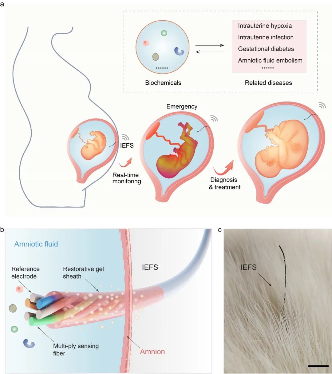
Figure 1. Schematic of the fiber sensor.
Interface Stabilization and Tissue Response
After implantation, the polymer gel swells within 15 minutes and seals the wound, adhering tightly to the amnion. The gel releases type I collagen, which promotes the conversion of amniotic epithelial cells to reparative amniotic mesenchymal-like cells. These cells secrete TGF-β-induced protein ig-h3 (TGF-βi), remodeling the periamniotic environment and facilitating wound healing. As evidence of preserved amnion integrity, levels of proinflammatory cytokines interleukin-6 (IL-6) and IL-1β, as well as amniotic fluid bacterial cultures, showed no statistical differences compared with blank controls. Matrix metalloproteinases MMP-8 and MMP-9, used as local stress indicators, were comparable to controls, and no additional amnion ruptures were observed during implantation (Figure 2).
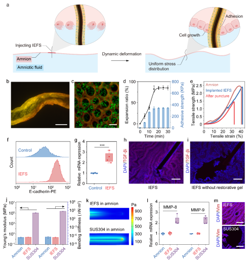
Figure 2. Stable device-tissue interface formed by the fiber sensor.
Biocompatibility
Because of the stable interface, the sensor showed good in vivo biocompatibility. No significant inflammatory cell infiltration was observed after implantation. Hematological and blood biochemistry indicators and the condition of major organs were consistent with blank controls. Fetal brain and limb development assessed by ultrasound showed no obvious differences, and gestational age and offspring survival in pregnant rats matched controls. Offspring body weight, brain development, and cognitive behavior showed no statistical differences compared with normal animals, supporting the sensor's biosafety (Figure 3).
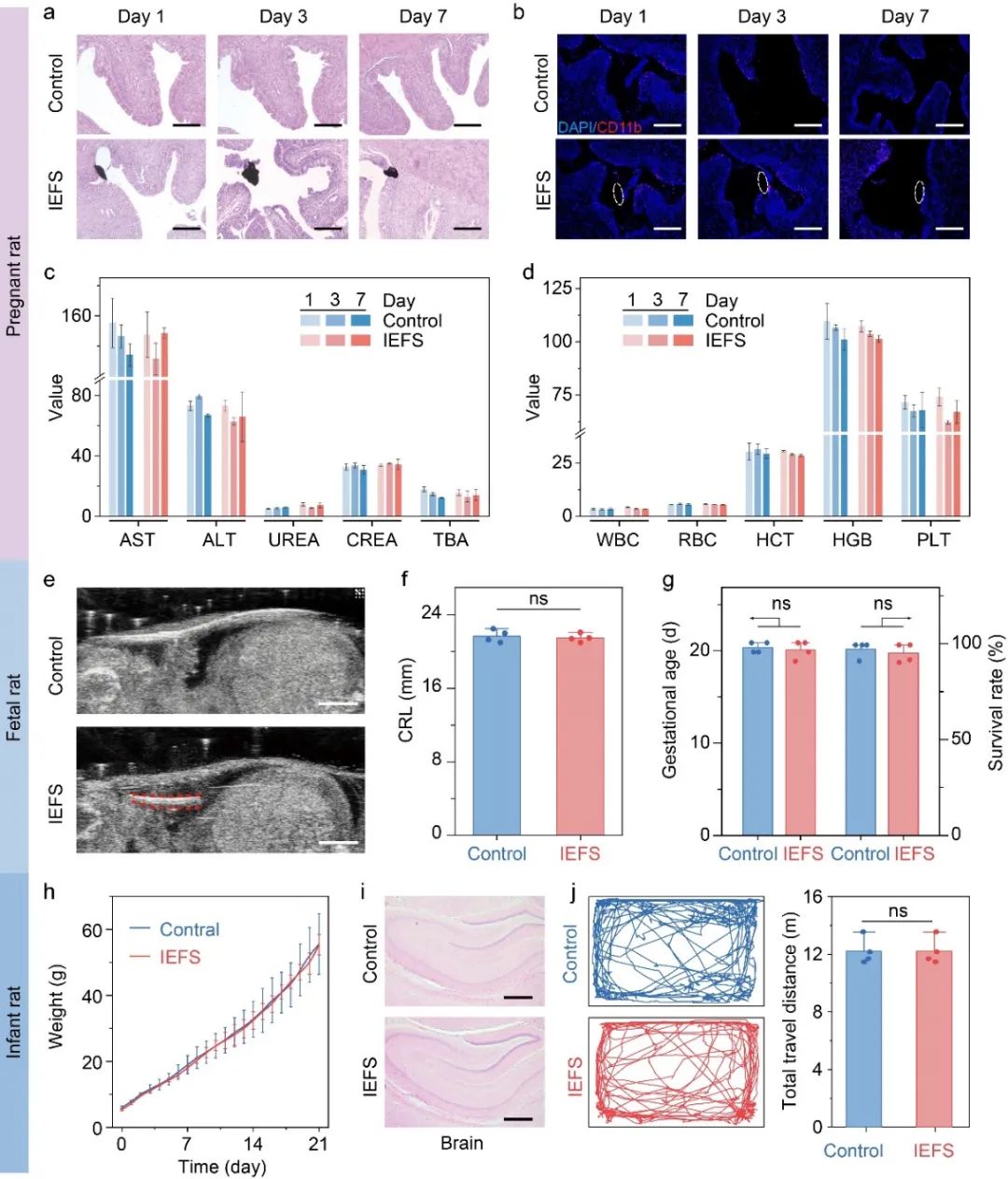
Figure 3. Biocompatibility assessment of the fiber sensor.
Response Performance
The sensor demonstrated high sensitivity and an appropriate response range for tracking dynamic fluctuations of relevant biochemical markers in amniotic fluid (Figure 4). After soaking in phosphate-buffered saline for 30 days, signal intensity remained stable. Biochemical signals detected in real time by the sensor matched the clinical gold standard (in vitro breakpoint measurement) with up to 98% accuracy. In simulated activities such as walking, shaking, jumping, and bending, the sensor response showed no significant interference.
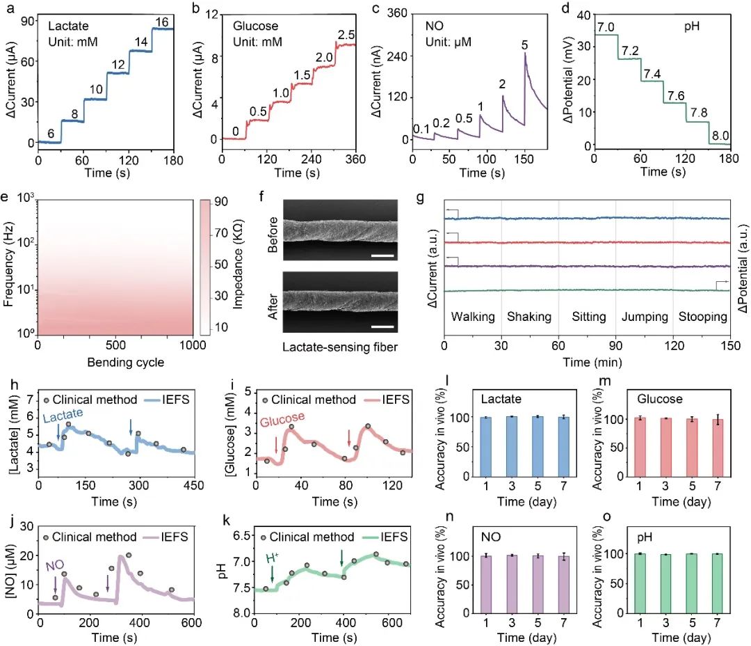
Figure 4. Response performance of the fiber sensor.
Integration and Application in Pregnancy Models
Integration of the fiber sensor with a flexible processing chip enabled early detection and warning of pregnancy disorders (Figure 5). The fiber sensor converts detected biochemical signals into electrical signals; a flexible chip collects and processes these signals and transmits data wirelessly via Bluetooth to a mobile device, enabling continuous real-time monitoring. In a rat intrauterine hypoxia model, amniotic fluid pH fell rapidly from 7.4 to 6.7 during acute hypoxia, while lactate concentration rose from 5.2 mM to 7.2 mM within 10 minutes. When lactate exceeded 7.2 mM, the sensor issued an early warning and appropriate interventions were applied. With this early warning, fetal survival reached 95%, close to unaffected levels. Without sensor-based warning, survival dropped to 13.3% after 1 hour, and pups exhibited marked reductions in brain erythropoietin and impaired brain development. These results indicate the sensor's potential value for early warning and improving fetal survival and development.
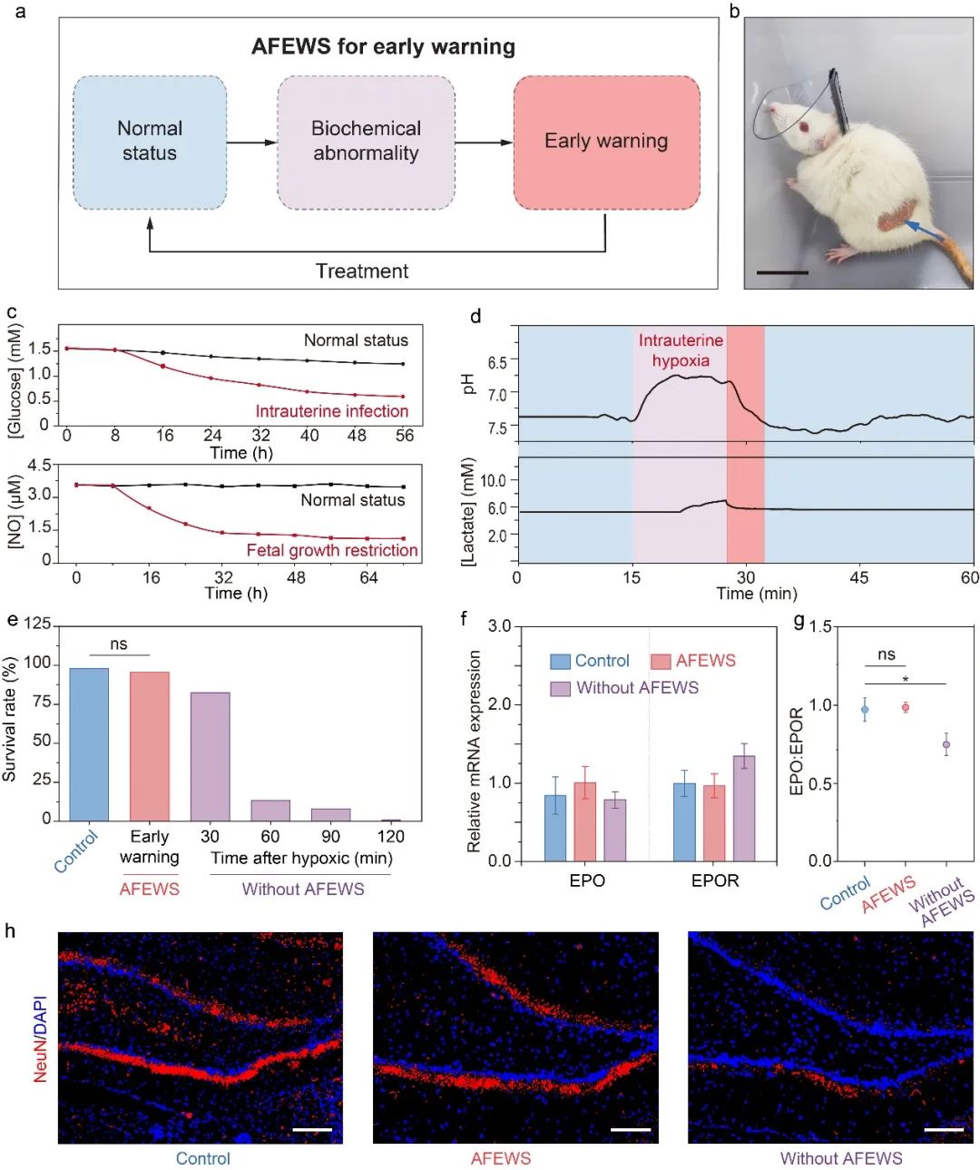
Figure 5. Application of the fiber sensor in pregnancy disorder models.
Conclusion
By designing a polymer gel coating that stabilizes the device-tissue interface, the work developed an interface-stabilized fiber sensor capable of continuous, real-time, and specific monitoring of multiple biochemical markers in amniotic fluid during pregnancy. This approach has potential value for early warning of pregnancy disorders and for understanding amniotic fluid biochemical dynamics. Expanding the range of monitored biomarkers could improve personalized prevention, diagnosis, and treatment of pregnancy-related conditions.